IEO: MOVING TOWARDS PRECISION MEDICINE WITH RADIOMICS, ONE OF THE MOST PROMISING RESEARCH AREAS

Precision medicine is a primary objective of contemporary medicine, aiming to tailor treatment to a patient’s specific features and illness. Radiomics is an expanding field that is emerging as a technology for personalized medicine, and it is currently one of the most intriguing areas of investigation.
The European Institute of Oncology was the first center in Italy to establish a Radiomics facility. Experts are diligently transforming the data acquired from radiological imaging tests into precise quantitative data, enhancing diagnostic accuracy and therapeutic effectiveness and thereby streamlining the clinical decision-making process.
Precision medicine is a primary objective of contemporary medicine, aiming to tailor treatment to a patient’s specific features and illness. Radiomics is an expanding field that is emerging as a technology for personalized medicine, and it is currently one of the most intriguing areas of investigation.
To understand what Radiomics is, it is necessary to start by saying that some tumors are characterized by molecular alterations, such as genomic alterations. Given that it is possible to define these alterations, it is generally necessary to have a sample of the neoplastic tissue, which is obtained by biopsies or invasive surgical interventions. Currently, however, imaging diagnostics can enable tissues to be characterized in a non-invasive manner and, in some cases, can enable the profound phenotypic differences that human tissues present to be visualized. Since tumors are heterogeneous in their volume and change over time, diagnostic images can provide a full view of the entire tumor and can be repeated over time to monitor the changes induced by therapies.
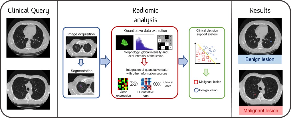
Through Radiomics, the medical images we know, obtained by CT, MRI or PET scans, are converted into numerical data. They are calculated, in very large numbers, by special calculation tools and their handling and analysis often require the use of advanced techniques, such as artificial intelligence methods, in order to manage so-called “big data”.
This huge wealth of numerical data, which could not possibly be processed by means of simple visual observation, defines many characteristics of the tumor and the surrounding environment, related, for example, to its shape, volume and tissue structure.
Using these techniques, it is possible to study any correlation between the data obtained from the images and the molecular and genomic characteristics of the tumor, with the final aim of extracting indications – directly from the images – regarding the aggressiveness of the disease, regarding the most commonly indicated therapies and regarding its response to treatment.
It is hoped that, in the near future, the data collected from radiological imaging tests will be converted into quantitative data and that this data will be used as a decision-making support to clinical practice, in order to improve diagnostic accuracy and prognostic power.
The European Institute of Oncology was the first center in Italy to establish a Radiomics facility. Experts are diligently transforming the data acquired from radiological imaging tests into precise quantitative data, enhancing diagnostic accuracy and therapeutic effectiveness and thereby streamlining the clinical decision-making process.


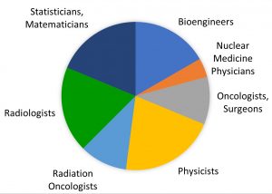 The IEO radiomics team includes medical doctors, physicists, engineers and statisticians. All members play an active role in radiomics projects.
 The IEO aims to become an international point of reference in the fight against cancer. For this reason, IEO International Office was set up in September 2013.